|
by Frank WATT and Barry HALLIWELL
 lectron microscopy is a mature technique for imaging structures and surfaces at resolutions well below the diffraction limit of light. An analogue of electron microscopy is proton microscopy, which uses high-energy protons. Although the technology for focusing protons is technologically more complex than for electrons (due to the much higher momentum of protons), proton microscopy has the unique feature of elemental mapping down to the parts-per-million level with high accuracy. The use of protons enables a suite of complementary techniques to be simultaneously utilised. Since the proton is the nucleus of a hydrogen atom, and is found in all nuclei, this suite of techniques is also known as nuclear microscopy (see box story). lectron microscopy is a mature technique for imaging structures and surfaces at resolutions well below the diffraction limit of light. An analogue of electron microscopy is proton microscopy, which uses high-energy protons. Although the technology for focusing protons is technologically more complex than for electrons (due to the much higher momentum of protons), proton microscopy has the unique feature of elemental mapping down to the parts-per-million level with high accuracy. The use of protons enables a suite of complementary techniques to be simultaneously utilised. Since the proton is the nucleus of a hydrogen atom, and is found in all nuclei, this suite of techniques is also known as nuclear microscopy (see box story).
 Nuclear microscopy can quantitatively map all elements in the periodic table as well as carrying out simultaneous structural imaging. The main drawback now is that high-energy proton focusing remains underdeveloped compared with the focusing of electrons. Current resolutions are limited to around 300nm for elemental analysis using Proton-Induced X-Ray Emission (PIXE) and Rutherford Backscattering Spectrometry (RBS) although a level of sub-100nm has been achieved for structural imaging (STIM). using PIXE and RBS showed an absence of aluminium, challenging the then-prevailing theory that aluminium toxicity might cause the disease. Nuclear microscopy can quantitatively map all elements in the periodic table as well as carrying out simultaneous structural imaging. The main drawback now is that high-energy proton focusing remains underdeveloped compared with the focusing of electrons. Current resolutions are limited to around 300nm for elemental analysis using Proton-Induced X-Ray Emission (PIXE) and Rutherford Backscattering Spectrometry (RBS) although a level of sub-100nm has been achieved for structural imaging (STIM). using PIXE and RBS showed an absence of aluminium, challenging the then-prevailing theory that aluminium toxicity might cause the disease.
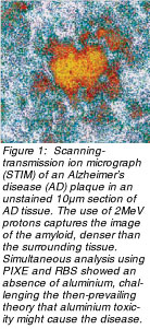
The instrument's ability to image structures in unstained tissue sections (Figure 1) while measuring trace elements gave rise to one early use of nuclear microscopy, described in a publication in Nature by Judith P Landsberg, Brendan McDonald, and Frank Watt (360:65-7,1992). They imaged amyloid plaques from brain sections of Alzheimer's disease (AD) victims and analysed them. The absence of aluminium in AD plaques from unstained tissue contrasted sharply with the high levels of aluminium observed in plaques previously identified and located using immunohistochemical stain. This work overrode the then-prevailing theory that aluminium might be a cause of AD and underlined the importance of developing stringent analytical protocols for microanalysis because elemental analysis of chemically fixed and stained tissue can lead to misinterpretation.
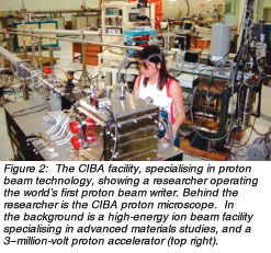 The Centre for Ion Beam Applications (CIBA) at the Department of Physics, National University of Singapore (NUS) houses a wide selection of sophisticated instrumentation. NUS is pioneering the use of high-energy protons for a wide variety of research applications (Figure 2), including proton microscopy. The CIBA proton microscope enables quantitative elemental mapping down to parts per million (ppm) levels and at spatial resolutions down to the cellular level and below. The Centre for Ion Beam Applications (CIBA) at the Department of Physics, National University of Singapore (NUS) houses a wide selection of sophisticated instrumentation. NUS is pioneering the use of high-energy protons for a wide variety of research applications (Figure 2), including proton microscopy. The CIBA proton microscope enables quantitative elemental mapping down to parts per million (ppm) levels and at spatial resolutions down to the cellular level and below.
Members of the CIBA group, in collaboration with colleagues from NUS's Departments of Biochemistry, Anatomy, and Pharmacology, are investigating the role of trace elements in such important degenerative diseases as Alzheimer's, Parkinson's, and atherosclerosis. Current research focuses on the role of iron, zinc, and copper in the development of such diseases.
Figure 3 shows an example of the potential of nuclear microscopy using protons, displaying the results of a proton scan across a cross-section of artery-wall tissue from a New Zealand White Rabbit fed a high cholesterol diet for a little less than two months. The high cholesterol induces atherosclerosis in the animal, and the atherosclerotic lesion shows clearly in the STIM image as a bulge on the side of the artery wall.
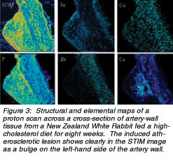 Simultaneous trace elemental mapping using PIXE and RBS led to four observations. Iron in the lesion rises, implying a probable increase in free radical reaction. Zinc, an element that may play a protective anti-atherosclerotic role, decreases. Copper also decreases, implying less likelihood of its involvement in free radical production. Finally, the presence of calcified deposits demonstrates the beginning of arterial hardening. Simultaneous trace elemental mapping using PIXE and RBS led to four observations. Iron in the lesion rises, implying a probable increase in free radical reaction. Zinc, an element that may play a protective anti-atherosclerotic role, decreases. Copper also decreases, implying less likelihood of its involvement in free radical production. Finally, the presence of calcified deposits demonstrates the beginning of arterial hardening.
Quantitative element analysis showed an increase of iron in early lesions, with an average of 90ppm compared with normal concentrations of 12ppm in the artery wall. The localised iron concentrations correlated strongly with the depth of the lesion in the artery wall, implying that local elevated concentrations of iron present in early lesions may accelerate atherogenesis in specific regions of the artery wall, as Watt, Barry Halliwell and their NUS collaborators found in a previous study (Free Radical Biology and Medicine 34(6):746-52, 2003). Unlike iron, zinc in the early lesion is depleted compared to the artery wall (about 50ppm compared with ~100ppm) and has a concentration inversely correlated with localised lesion depth, implying that zinc may have an anti-atherosclerotic effect. Further work by the researchers feeding rabbits a zinc-enriched diet has supported this result (Free Radical Biology and Medicine. In Press (2006)).
Copper levels in general are much lower and nearer the levels of detection for PIXE, with depletion in the lesion (~2ppm) compared with the smooth muscle cells in the artery walls (~4ppm). In newly developed lesions, scientists observed depleted calcium concentrations (~450ppm) compared with the adjacent healthy wall (~575ppm) whereas in more mature lesions, they saw mineral deposition form first in the lesion/artery wall interface and subsequently in the lesion. The results have implications for further study of the effects of trace elements on the progression of degenerative diseases.
Nuclear microscopy is in its infancy, and CIBA remains one of very few facilities worldwide employing this method for biomedical research. It offers a unique and useful tool in biomedicine -- that of simultaneous imaging and quantitative microanalysis down to the sub-cellular level. This fresh approach will provide valuable insight into the mechanisms driving the occurrence of certain diseases and perhaps help researchers in their quest for possible intervention.
|
Nuclear Microscopy Techniques
Particle Induced X-ray Emission (PIXE)
When a fast-moving proton collides with matter, a high probability exists that atoms in the material will eject inner-core electrons, creating vacancies in the inner-core electron shells. Electrons from the outer shells will fill these vacancies almost immediately while releasing X-ray photons with an energy characteristic of the parent atoms. The detection and subsequent identification of these proton-induced X-rays allow elemental analysis of the sample. Focusing the proton beam down to sub-micron spot sizes and scanning the beam across a sample can construct elemental maps down to the parts-per-million level.
Scanning Transmission Ion Microscopy (STIM)
If the sample is thin enough (such as a biological tissue of less than 20μm), then the protons have sufficient energy to pass through the sample, losing power as they go. Researchers measuring the energy loss of the transmitted protons can construct a density map of the sample.
Rutherford Backscattering Spectrometry (RBS)
Although most protons will interact with the sample atoms via proton/electron collision, a small fraction will collide with the nuclei of the sample atoms. In this case, the rebounding protons will lose energy that depends on the mass of the nucleus. Since protons backscattered from the lighter nuclei lose more energy, measurement of the energy of backscattered protons allows quantitative assessment of the light elements (for example, carbon and oxygen) in the sample.
|
 Click here to download the full issue for USD 6.50 Click here to download the full issue for USD 6.50
|