|
by Dr Wieslaw L Nowinski and A Thirunavuukarasuu
 lthough brain atlases in printed form have been available for five decades - and computerised atlases were introduced for use in neurosurgery in the 1970s - the practical impact of all these activities on daily clinical practice was low. This situation promises to change with the availability of a new electronic brain atlas developed by a group of Singapore-based scientists. lthough brain atlases in printed form have been available for five decades - and computerised atlases were introduced for use in neurosurgery in the 1970s - the practical impact of all these activities on daily clinical practice was low. This situation promises to change with the availability of a new electronic brain atlas developed by a group of Singapore-based scientists.
The Biomedical Imaging Lab (BIL) at the Laboratories for Information Technology (formerly Kent Ridge Digital Laboratories) has constructed a novel electronic brain atlas database called the Cerefy brain atlas database and established it as a standard in neurosciences. This database was derived from several printed atlases well known to clinicians and researchers and used extensively by them.
The most difficult task of the Cerefy project was building the bridge between information technology and neurosciences. This work is the most detailed brain atlas database containing information on different parts of the brain, such as gross anatomy, subcortical structures, brain connections, and cortical patterns. To create the Cerefy atlas, scientists at BIL have registered the multiple atlases constructed in such a manner that they correspond to one another seamlessly in three-dimensional (3D) space. From this database, they built 3D models of subcortical structures and cortical areas to facilitate the understanding of spatial brain relationships. The researchers have developed and patented a fast method to match the atlas to data. They have also successfully commercialised solutions for surgery of movement disorders such as Parkinson's disease as well as neuroradiology, human brain mapping, and neuroeducation.
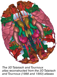
The process of atlas construction was complicated and laborious, demanding high accuracy and extensive anatomical knowledge. The original printed atlases were digitised, enhanced, extended, segmented, labelled, aligned, and organised into atlas volumes. The anatomical index has about 1,000 fully segmented and labelled structures per hemisphere and over 400 cortical patterns. From the database, researchers constructed 3D atlases and mutually co-registered them with the 2D atlases.
The research has resulted in the filing of three international patents. One of the patents already granted covers a novel method of annotating radiological images with anatomical names. The user reads the radiological images in the standard Digital Imaging and Communications in Medicine (Dicom) format, which allows medical images to be exchanged, defines the regions of interest and annotates them, then segments and labels the structures within these defined regions, using the brain atlas. The annotated and labelled images are saved in the Dicom or XML format.
The images enhanced in this way can be accessed in Picture Archiving and Communication Systems (PACS), commonly used in hospitals or in Web-based applications for further processing.
Brain Atlas Applications
Starting from the Cerefy brain atlas database, the team has developed a set of atlas-based applications for neurosurgery, brain mapping, neuroradiology, and neuroeducation. Some of the ideas and input come from collaboration, particularly in functional neurosurgery, with local and international medical organisations such as Singapore's Tan Tock Seng Hospital, its National Neuroscience Institute, Johns Hopkins University/Hospital, Massachusetts General Hospital, and the Mayo Clinic.
BIL scientists have licensed products and technology to several leading global companies. In addition, they have created nine products distributed worldwide, including four low-cost CD-ROMs, and two plug-in libraries, namely Electronic Brain Atlas Library and Brain Atlas Geometrical Models. These libraries have become the standard in functional neurosurgery to treat Parkinson's disease patients and have already been integrated with surgical systems of major image-guided surgery companies such as Medtronic/Sofamor-Danek, BrainLAB, and Elekta.
The brain atlas is particularly helpful in surgery of movement disorders to identify critical structures usually not clearly visible on the scan. The use of the atlas significantly lowers the surgical cost by reducing the duration of surgery and the number of electrodes inserted into the brain. It also allows for rapid surgical planning, increases the accuracy of surgery, decreases potential surgical complications, lowers the invasiveness of the surgical procedure, increases the neurosurgeon's confidence, and reduces the time medical students spend learning neuroanatomy and scanning interpretation.
For more information contact Dr Wieslaw Nowinski at wieslaw@lit.org.sg or A Thirunavuukarasuu at thiru@lit.org.sg. Also check out www.cerefy.com.
|